
Anterior lower-leg muscles
Tibialis anterior
Musculus tibialis anterior
Your front shin muscle lifts the forefoot and controls how it lowers — helping your foot clear the ground and land smoothly as you walk.
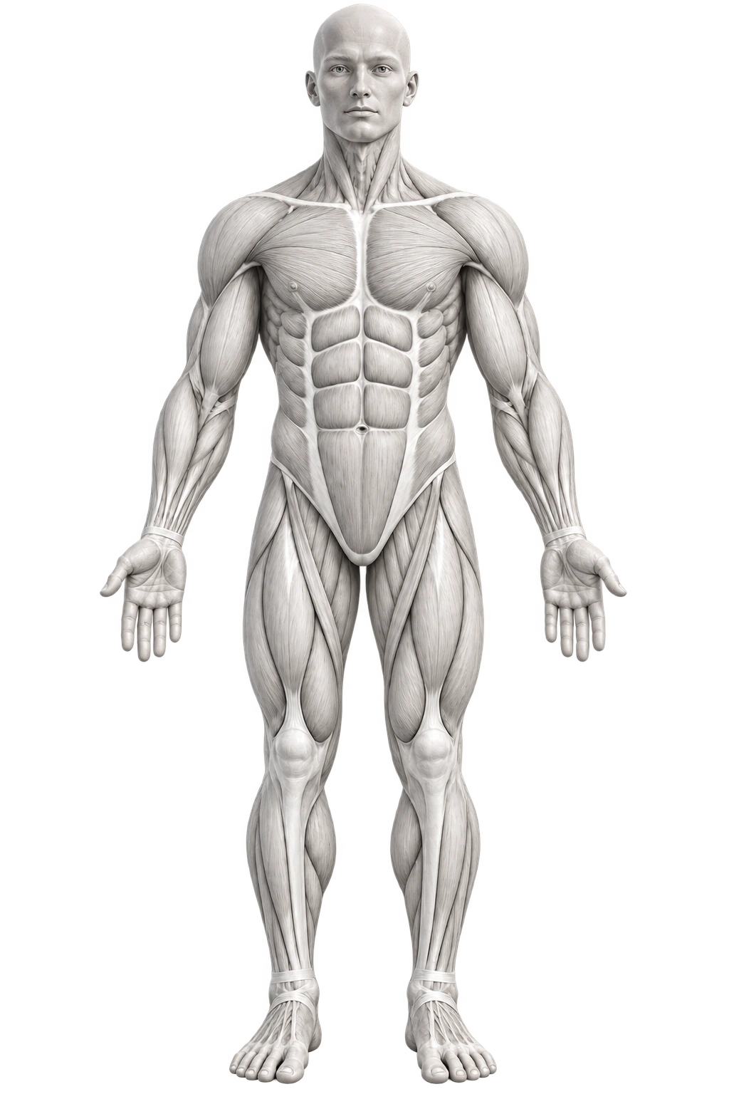
At a glance
- Region
- Front lower leg
- Movement pattern
- Forefoot lifting and controlled lowering
- Joint
- Ankle and foot joints
Exercises
Exercises for the tibialis anterior
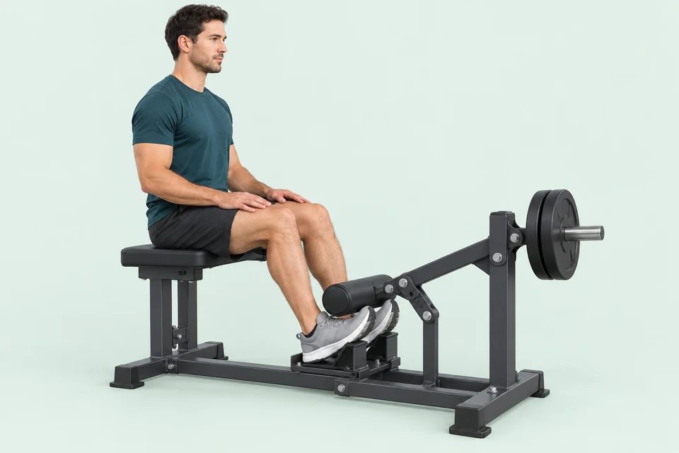
Dorsiflexion machine · Upper-foot pad
Tibialis raise machine
Raise the forefeet toward the shins against a padded lever while the heels remain supported.
View technique
No equipment · Chair or bench
Seated toe raise
Lift the forefeet while seated, keeping both heels on the floor. A simple way to practise ankle dorsiflexion.
View technique
No equipment · Wall
Wall tibialis raise
Support your back against a wall and lift the forefeet. Your heels remain the contact point with the floor.
View technique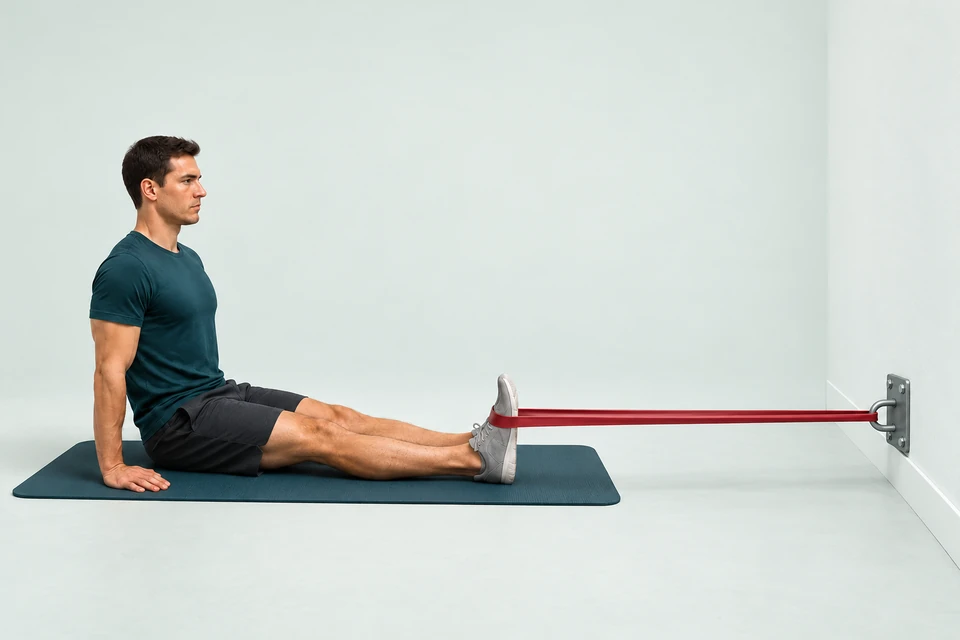
Resistance band · Secure anchor
Banded ankle dorsiflexion
Pull the forefoot toward you against a band anchored in front. Train one side at a time.
View techniqueExercise suggestions for this muscle are being prepared.
Understand the muscle
From the shin to the inner foot
One muscle belly continues into a long tendon across the front of the ankle. Its attachment on the inner foot allows it to lift the forefoot and help turn the sole inward.
Alongside the shin
Muscle belly
Venter musculi tibialis anteriorisBegins at the upper outer tibia and the membrane between tibia and fibula. This fleshy section generates the pulling force.
Exercises for this region
Across the front of the ankle
Long tendon
Tendo musculi tibialis anteriorisContinues from the lower part of the muscle and passes beneath retaining bands at the ankle, transmitting force to the foot.
This is an attachment or connective-tissue structure, not a separate contractile muscle region. The exercises for the whole muscle train the associated movement.
Medial midfoot
Inner foot attachment
Os cuneiforme mediale et os metatarsale primumAttaches to the medial cuneiform and the base of the first metatarsal. It acts on the foot rather than attaching to the toes.
This is an attachment or connective-tissue structure, not a separate contractile muscle region. The exercises for the whole muscle train the associated movement.
The listed exercises involve this region alongside other parts of the muscle. A muscle head or deep portion is not necessarily trained in isolation.
Simply explained
What does the muscle do?
The tibialis anterior moves the foot at the ankle. It lifts the forefoot, helps turn the sole inward and slows the foot's descent after the heel touches down.
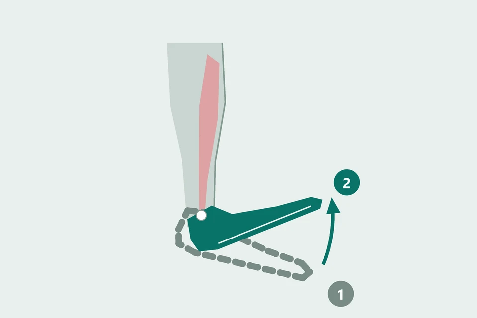
Lift the forefoot
Ankle dorsiflexion
The foot moves toward the shin at the ankle. During the swing phase of a step, this helps the forefoot clear the floor.
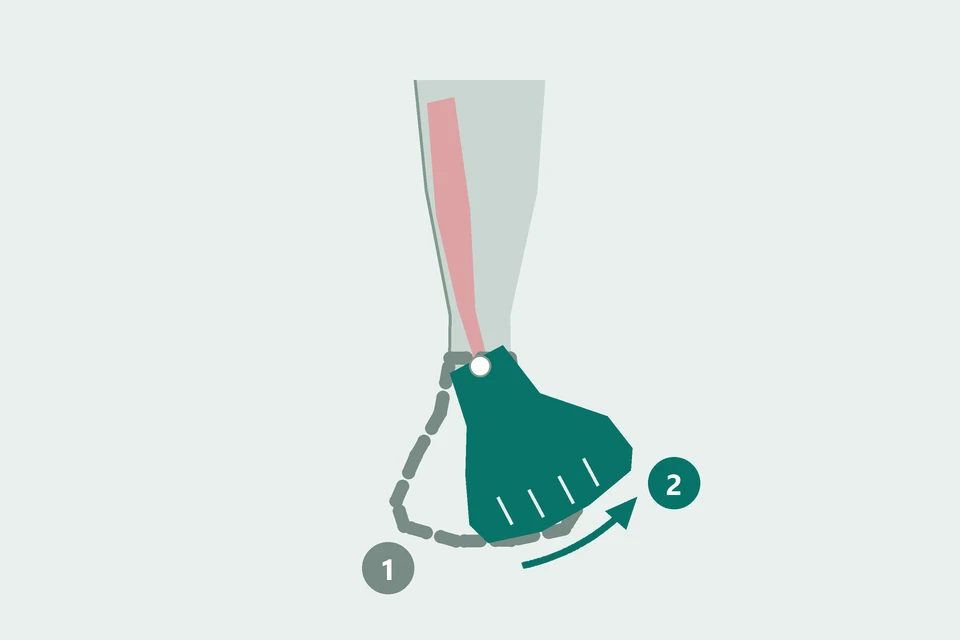
Turn the sole inward
Foot inversion
The tibialis anterior helps lift the inner edge of the foot and turn the sole inward. It shares this action with the tibialis posterior.
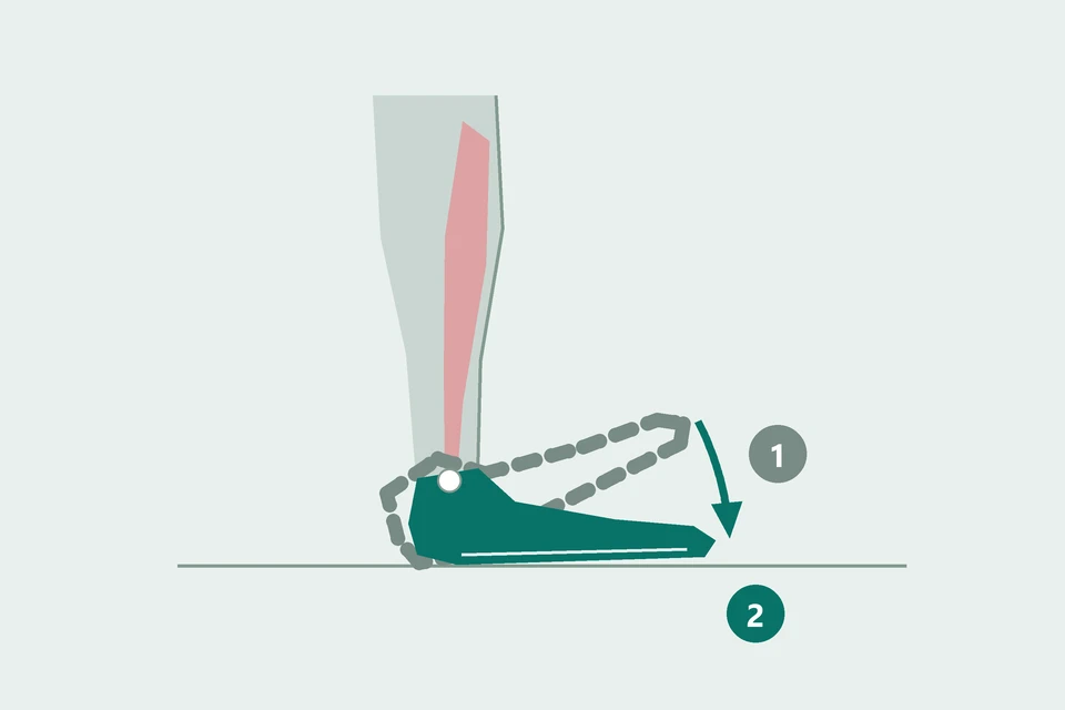
Lower the foot gently
Eccentric control
After the heel touches down during walking, the muscle produces force as it lengthens. This slows the forefoot's descent to the ground.
Often works with
Sources & illustrations
Anatomy and technique: the references below. Region boundaries and movement diagrams are schematic. Exercise images are AI-generated illustrations.